he testis, an essential organ of the male reproductive system, plays a central role in the production of sperm and the synthesis of testosterone, the primary male sex hormone. Understanding the anatomy of the testis is crucial for comprehending its functions and the various processes involved in male fertility and reproductive health. In this informative guide, we will explore the intricate anatomy of the testis, including its structure, function, and significance in male physiology.
Anatomy of testis

Anatomy of testis mean the testis is a paired, oval-shaped organ located within the scrotum, the external sac that hangs below the penis. Each testis is approximately 4 to 5 centimetres in length and is suspended within the scrotum by the spermatic cord, which contains blood vessels, nerves, and the vas deferens, tube that travels from the testis to the urethra to carry germs.
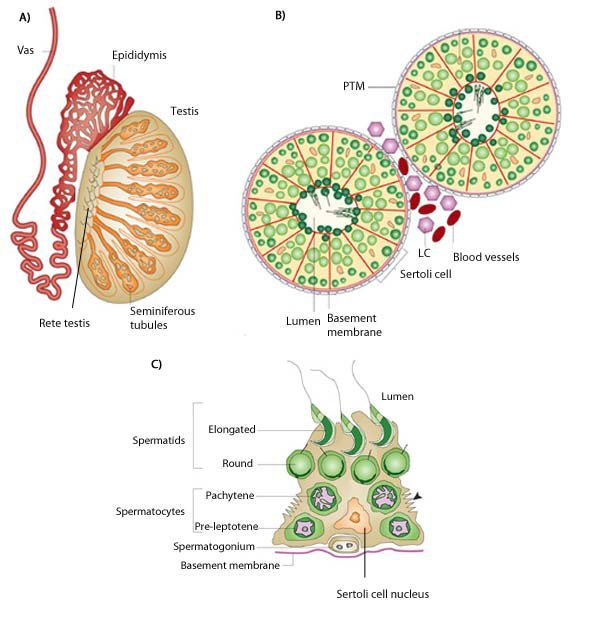
The outer surface of the testis is covered by a dense fibrous capsule known as the tunica albuginea, which helps maintain the structural integrity of the organ. Inside the testis, the tunica albuginea extends inward, dividing the testis into lobules or compartments, each containing one to four highly coiled tubules known as seminiferous tubules.
Seminiferous Tubules
The seminiferous tubules are the functional units of the testis responsible for sperm production, a process known as spermatogenesis. These tubules are lined with specialized cells called Sertoli cells, which provide structural support and nourishment to developing sperm cells, or spermatids.
Spermatogenesis begins at the periphery of the seminiferous tubules, where germ cells, known as spermatogonia, undergo a series of mitotic divisions to produce primary spermatocytes. These primary spermatocytes then undergo two rounds of meiotic divisions, resulting in the formation of haploid spermatids.
As spermatids mature, they undergo a process called spermiogenesis, during which they undergo structural and morphological changes to develop into fully mature spermatozoa or sperm cells. Once mature, sperm cells are released into the lumen of the seminiferous tubules and transported to the epididymis for further maturation and storage.
Interstitial Tissue
Anatomy of testis in addition to seminiferous tubules, the Anatomy of testis contains interstitial tissue, also known as the interstitial or Leydig cells, located in the spaces between the tubules. Leydig cells are responsible for producing testosterone, the primary male sex hormone essential for the development and maintenance of male reproductive tissues and secondary sexual characteristics.
Testosterone production is regulated by luteinizing hormone (LH), a hormone released by the pituitary gland in response to signals from the hypothalamus. LH stimulates Leydig cells to produce testosterone, which then exerts its effects on target tissues, including the seminiferous tubules, accessory reproductive glands, and secondary sexual organs.
Blood Supply and Innervation
The testis receives its blood supply from the testicular arteries, which arise from the abdominal aorta and descend into the scrotum alongside the spermatic cord. Blood is drained from theAnatomy of testis by the testicular veins, which form the testicular plexus and ultimately converge to form the left and right testicular veins.
The testis is also innervated by the autonomic nervous system, with sympathetic and parasympathetic fibres originating from the spinal cord and travelling along the spermatic cord to provide sensory and motor innervation to the testicular tissues. These nerves play a role in regulating blood flow, temperature, and contractile activity within the testis.
The function or Anatomy of testis
The primary functions of the testis include:
Spermatogenesis:
The production of sperm cells through a complex series of cell divisions and maturation processes within the seminiferous tubules.
Testosterone Production:
The synthesis of testosterone by Leydig cells, which is essential for the development and maintenance of male reproductive tissues, secondary sexual characteristics, and overall health.
Regulation of Reproductive Function:
in Anatomy of testis the testis plays a central role in regulating reproductive function through the production of sperm and testosterone, which influence fertility, sexual development, and libido.
Clinical Considerations
before we Understanding the Anatomy of testis and function of the testis is crucial for diagnosing and managing various conditions affecting male reproductive health. Common disorders and conditions involving the testis include.Anatomy of testis
Testicular Torsion:
A medical emergency is characterized by the twisting of the spermatic cord, which can disrupt blood flow to the testis and lead to severe pain and potential tissue damage.
Testicular Trauma:
Injuries to the testis result from direct trauma or blunt force, which can cause pain, swelling, and potential damage to testicular tissues.
Cryptorchidism:
A congenital condition in which one or both testes fail to descend into the scrotum, increasing the risk of infertility and testicular cancer.
Testicular Cancer:
Malignant tumors arise from the cells of the testis, which may present as painless testicular lumps, swelling, or changes in testicular size.
Conclusion
The testis is a complex and vital organ of the male reproductive system, responsible for sperm production, testosterone synthesis, and the regulation of reproductive function. Understanding the anatomy and function of the testis is essential for comprehending male reproductive health and diagnosing and managing various conditions affecting testicular function. By maintaining awareness of the structure and function of the testis, healthcare professionals can provide effective care and support for patients experiencing issues related to male fertility and reproductive health.

